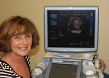
Director: Sally Hill MSc
ULTRASOUND SCANS
Diagnostic Ultrasound Services have state of the art ultrasound equipment providing patients and referring doctors with a comprehensive range of ultrasound scans. Our sonographers and radiologists are highly qualified.
WHAT IS AN ULTRASOUND SCAN?
Ultrasound is a painless test involving the use of sound waves. Ultrasound scanners use high-frequency sound waves to create an image of the inside of the body. Ultrasounds are used to look for changes in tissue in organs. There are no harmful effects.
WHAT ARE ULTRASOUNDS USED FOR?
- Examining the kidney, bladder and prostate gland to identify abnormalities
- Diagnosing tumours, gallstones and cysts in organs such as the liver and pancreas
- Diagnosing muscle and tendon tears in musculo-skeletal conditions.
- Visualising the uterus and ovaries to check for ovarian cysts/fibroids/endometriosis/tumours and polycystic ovaries.
- Used very commonly in pregnancy to see if the baby has developed normally and whether it is growing well.
WHAT HAPPENS IN AN ULTRASOUND?
You will be shown to the ultrasound room and asked to lie on the ultrasound couch on your back while the scans are performed. A clear lubricating jelly is applied to the area being imaged to help the transducer make secure contact with the body. This eliminates air pockets between the transducer and the skin resulting in clearer images. The sonographer then gently presses the transducer firmly against the skin and sweeps it over the area of interest until the desired images are captured. A pelvic scan to see the uterus and ovaries is better seen using a trans-vaginal probe although we can do a tummy scan if necessary.
The ultrasound probe directs a stream of high frequency sound waves in to the body. The sound waves are reflected off the internal organs and structures in the body. The reflected ultrasound waves are detected by the transducer and used to create an image of the organs and structures. Ultrasound is captured in real time and is constantly updating so that you can see movement, such as the valves of a heart opening and closing.
Ultrasound is painless and will take typically between 10 – 25 minutes. A written report is available immediately after your scan and this will be sent to your GP or referring doctor.
WHAT ARE THE BENEFITS OF ULTRASOUND?
- Painless and non-invasive
- Can visualise movement and function and therefore can examine blood vessels and blood flow to different organs
- Does not use x-rays or any other type of ionising radiation to produce an image
- Gives a clear image of soft tissues that may not show up well on x-rays
- Causes no health problems and may be repeated as often as necessary
HOW SHOULD I PREPARE FOR AN ULTRASOUND?
There may be special preparation required for an ultrasound depending on the area of the body being imaged. You may need to fast for a few hours before certain ultrasounds, or required to have a full bladder.
You will be given any specific instructions when you book your appointment.

