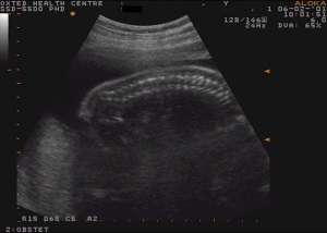
Fetal anomaly scan: 21 – 23 weeks (Second trimester)
The fetus will now measure about 10 inches (25cms) in length and the purpose of this scan is to examine the anatomy, ensure normal growth and check the placental position. Uterine blood flow studies can be performed when indicated. Cervical length can be assessed to evaluate the risk of pre-term delivery

What do we look for?
The structures examined include the brain, spine, heart, kidneys and limbs. Sometimes the fetus is in a position which makes scanning difficult and it is quite normal to be asked to return for a second appointment to complete the examination. Generally the image is not as clear in larger women and again a second appointment may be necessary.
Approximately 90% of significant abnormalities will be detected.
What if a problem is detected ?
Not all abnormalities are life threatening.
For example, sometimes there is an excess of urine within the fetal kidneys, which can be monitored by further scans. Most kidneys return to normal by the end of the pregnancy, but early detection reduces the incidence of childhood kidney infections and obstructions. If a more serious abnormality is suspected, a second opinion at a specialist centre may be arranged to discuss the best management of the pregnancy.
Remember that if there are any major problems we should be able to pick them up, but it is important to realise that not all abnormalities can be diagnosed – we recommend that this scan is best done between 20 – 22 weeks.
Doppler Blood Flow Measurement
Doppler blood flow measurements of the uterine artery to predict the later development of IUGR and pre-eclampsia.

